A sudden three-legged hop mid-walk. The leg comes back down a few seconds later, and the dog carries on like nothing happened. Owners call it "the skip" and post it on TikTok like it's adorable. It isn't. It's the kneecap leaving its track and grinding cartilage off the bone every time it does.
I. What Patellar Luxation Is
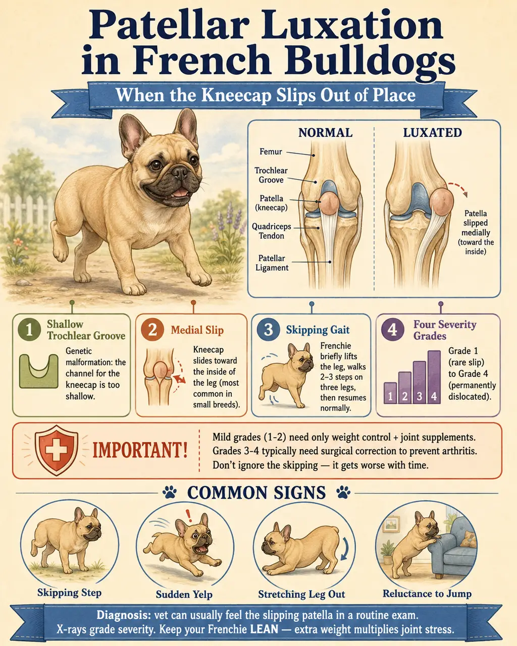
The kneecap (patella) pops out of the groove at the bottom of the femur. Instead of tracking smoothly up and down as the leg bends and straightens, the kneecap slides off — usually toward the inside (medial patellar luxation, or MPL).
Each dislocation grinds cartilage. Over time the groove itself wears flat, and the dislocation gets worse and worse.
II. Why Frenchies Are Prone
- Small breed size — patellar luxation is most common in small breeds (Poodles, Pomeranians, Yorkies, Frenchies).
- Structural skeletal abnormalities — bowed femurs, shallow trochlear groove (the channel the kneecap rides in), rotated tibias from chondrodystrophy.
Females are more predisposed than males. Neutered dogs have approximately 3× the odds of developing patellar luxation.
The combination of broad squat frame, short legs, and chondrodystrophic bone is a textbook case.
III. The Four Grades
| Grade | Description |
|---|---|
| Grade 1 | Patella stays in place during normal use. Vet can manually push it out, and it pops back when released. Often asymptomatic. |
| Grade 2 | Spontaneously dislocates during activity (the classic "skipping" gait). Returns when the dog extends and rotates the leg. Most common in Frenchies. |
| Grade 3 | Patella OUT most of the time. Vet can push it back, but it pops out again. Visible bow-leggedness. |
| Grade 4 | Permanently dislocated, cannot be manually repositioned. Crouched, severely bow-legged gait. Often cannot fully extend the leg. |
IV. Symptoms Owners Notice First
- The "skip" — sudden three-legged hop mid-walk, back to normal in a few steps.
- Sudden, brief lameness that resolves quickly.
- Holding leg up for a few seconds, then putting it back down and resuming.
- Crackling or popping sound at the knee.
- Reluctance to jump or run.
- Sitting with leg out to the side instead of tucked under.
- Bow-legged stance (Grade 3–4).
- Stiff gait after rest.
We thought it was a quirky little hop. Six months later the vet said it was Grade 3 in both knees and we needed surgery on each.
V. How It Gets Diagnosed
- Physical exam — the vet feels the patella sliding in and out and assigns a grade. This is the primary diagnostic tool. No special equipment needed.
- X-rays — confirm severity, look for arthritis, check concurrent issues (cruciate damage, malformed femur).
- CT — surgical planning in severe cases.
VI. Treatment by Grade
| Grade | Treatment |
|---|---|
| 1 | No surgery. Monitor, weight management, supplements, avoid high-impact activity. |
| 2 | Borderline. Many vets recommend surgery sooner to prevent progression and concurrent CCL tear. Conservative management can work but carries arthritis risk. |
| 3 | Surgery recommended. Conservative management leads to severe arthritis. |
| 4 | Surgery required to restore meaningful function. |
VII. Surgery — What's Involved
Procedures (often combined)
- Trochleoplasty — deepens the groove the patella sits in.
- Tibial tuberosity transposition — relocates the bony attachment point on the shin to realign the pull of the kneecap.
- Soft tissue rebalancing — releases the tight side and tightens the loose side of the joint capsule.
Cost & outcomes
- Cost: $1,500–$5,000 per knee (Grade 2–3 typically $2,000–$3,000; Grade 4 or specialty hospitals can reach $5,000).
- Success rate: ~95% regain good knee function and normal lives.
- Recovery: 6–8 weeks of restricted activity, leashed walks only, gradual return.
VIII. Bilateral — The Other Knee
Approximately 50% of cases are bilateral — both knees affected. Owners often notice one bad knee and only later realize the other has been compensating silently.
If your Frenchie has Grade 2+ on one side, the other side is at high risk and should be examined. Bilateral surgery is often planned with a gap of weeks or months between knees, but knowing the status of both is essential.
IX. Common Owner Mistakes
- Treating the skip as cute behavior — every dislocation grinds cartilage off permanently.
- Waiting "to see if it gets worse" — Grade 2 progressing to Grade 3 means a much harder surgery.
- Allowing jumping — the biggest preventable trigger.
- Not getting the OTHER knee checked after diagnosis.
- Skipping post-op rehab — surgery is only half the recovery; PT determines outcome.
- Letting weight creep up — Frenchies are food-motivated, and obesity multiplies joint forces.
References
- Patellar Luxation in Dogs (Canadian Vet Journal). PMC6055913
- VCA — Luxating Patella in Dogs. VCAhospitals.com
- VET4BULLDOG — MPL in Bulldogs. Vet4Bulldog.com
- Recovery After MPL Surgery in Dogs. SustainableVet.org
- PetMD — Luxating Patella in Dogs. PetMD.com